Specialists of office of radiodiagnosis, doctors and average health workers of the highest and first categories, conduct researches with application of beam methods of visualization in a computer tomography, radiological and ultrasonic diagnostics.
 The office has the magnetic and resonance tomograph (MRT).
The MRT method is based not on the types of radiation (for example, the ionizing radiation) used for a roentgen or the computer tomography (CT), and on a potent, constant magnetic field, quickly changing local magnetic fields and radio-frequency energy. In MRT the express equipment which creates very sharp images is used. According to the obtained data the doctor can distinguish the slightest changes or violations. Use of MRT in diagnostics allows to establish the diagnosis in short terms and to carry out differential diagnostics at diseases of a brain, a locomotorium and internals. One more plus of a method – it safe and absolutely painless. Only the people having claustrophobia can feel unpleasant feelings, MRT can be contraindicated to them.

The office is equipped with the following equipment of Siemens:
a) The computer tomograph – the 16-slice spiral SOMATOM EMOTION tomograph
On KT researches of the following areas are carried out:
- brain;
- skull bones;
- okolonosovy adnexal bosoms;
- bodies of a thorax;
- abdominal organs and small pelvis;
- backbone;
- joints;
As indications to a research on the computer tomograph serve existence or suspicions on existence of traumatic damages, anomalies, new growths, inflammatory processes and other pathological states. The spiral computer tomography allows to reveal pathological changes in the studied bodies with higher precision.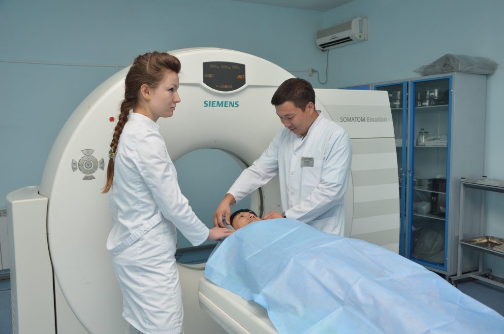
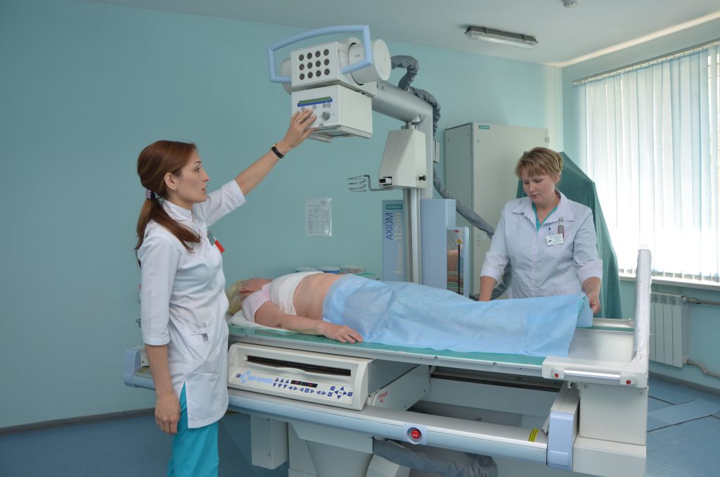
b) The radiological office of the accident ward is equipped with the stationary device MULTIXPROP on two workplaces:
- R-grafiya
- R-tomography
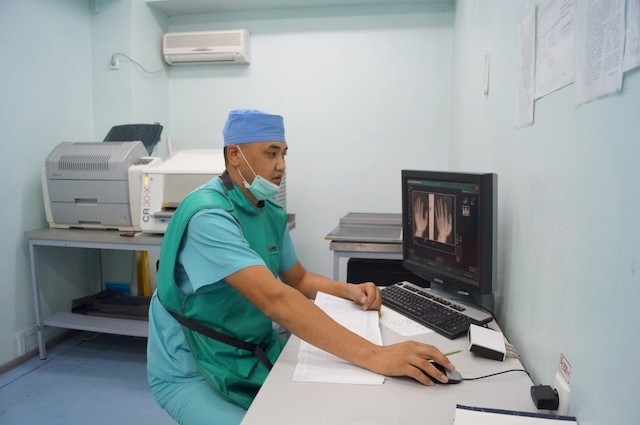
The digital roentgenography reduces time of inspection and manifestation of pictures.
c) The R-device AXIONICONOS R-200 on three workplaces:
- R-grafiya;
- R-skopiya;
- R-the tomography – examines inpatients
All types of X-ray analyses are carried out:
- a roentgenoscopy (exposures to radiation at diseases of internals): gullet, stomach, 12-tiperstny gut, large intestine, thorax, heart;
- roentgenography – pictures at the most various diseases and damages;
In time carrying out specialist research (excretory urography)
(With – an arch) “ARKADICVARIC” allows to conduct examination in all projections inspection of operational patients.
Under monitoring of the device the transpedikulyarny metalosteosynthesis is carried out.
Except stationary X-ray apparatuses there are 3 mobile X-ray apparatuses of POLYMOBIL which serve operational patients and patients on places.
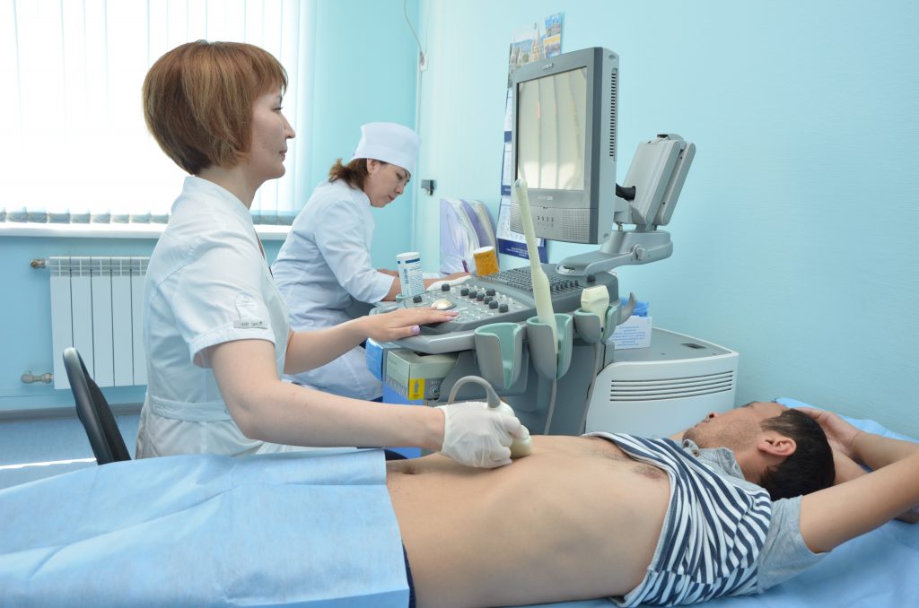
4 ultrasonography of the device ACUSON X-300 and 1 ultrasonography device Maylab 50:
- Ultrasonography of abdominal organs and retroperitoneal space: liver and gall bladder, pancreas, spleen, kidneys and adrenal glands, limfuzl;
- Ultrasonography of a small pelvis (bladder, prostate gland, uterus and ovaries);
- Ultrasonography of mammary glands;
- Ultrasonography of a thyroid gland;
- Ultrasonography of the surface lymph nodes vessels (arteries and veins) and others
Preparation for ultrasonography
Ultrasonography of a liver, gall bladder, pancreas, kidneys
In 3 days prior to a research to exclude black bread, crude vegetables, fruit, peas, haricot, potatoes, cabbage in any kind, milk and dairy products, sweets. It is possible: low-fat meat, cheese, white loaf (it is better dried), kissels, porridges on water. For elimination of the raised aerogenesis to accept one of the specified medicines: “Festalum” — 1 t. 3 times a day, “Mezim-forte”, “Pancreatinum” – 1-2 t. during food, Espumizan – 2 capsules 3 times a day. The dinner is mild, no later than 18 clocks: white loaf (crackers) with tea, porridge on water.
Not to have breakfast, not to smoke, not to drink water in the morning!
Ultrasonography of a bladder, prostate, uterus
The diet is described above, on a research to be with the complete bladder (in 2 hours prior to a research to drink 1,0 – 1.5 liters of water without gas).
For all specified researches intestines have to be emptied.
At itself to have a towel (diaper), removable footwear or boot covers
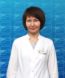
 The manager of office of radiodiagnosis is Adenova Gulzhan Moldabekovna